Staphylococcus Vs. Streptococcus.
A Comprehensive Analysis. Comparison and Contrast.
Author:
Dr.B.Fidanoski, DMD
I. Mutual Characteristics: Both genera are Gram positive and have the similar spherical cell shape, therefore there are called cocci (in Greek coccus means granule).
II. Visual differentiation between Staphylococci and Streptococci by cellular arrangement:
When performing laboratory analysis, after Gram staining, first thing we should do is put a piece of a colony under microscope and observe cellular arrangement of the bacteria. Both staphylococci and streptococci have round, spherical cell shape, but the arrangement of cells is different due to a different binary fission. Streptococci form a chain of round cells, because their division occurs in one linear direction, whereas staphylococci divide in various directions forming grape-like clusters.

III. Biochemical differentiation between Staphylococci and Streptococci with CATALASE test:
The main criterion for differentiation between Staphylococcus and Streptococcus genera is the catalase test. Staphylococci are catalase positive whereas Streptococci are Catalase negative. Catalase is an enzyme used by bacteria to induce the reaction of reduction of hydrogen peroxide into water and oxygen.
III-A. Biochemical differentiation among Staphylococci:
There
are at least three Staphylococci species of clinical importance:
1. Staphylococcus aureus is the most pathogenic for humans
2. Staphylococcus epidermidis, which is part of the normal flora and is
of low pathogenicity, and
3. Staphylococcus saprophyticus which can cause urinary tract
infections, especially in sexually active young women.
1.Differentiation
between Staphylococcus aureus and other Staphylococci:
- COAGULASE test which is
positive for Staphylococcus aureus (generally accepted criterion for the
identification) and negative for all other Staphylococci. Coagulase is an enzyme
used by S.aureus to induce coagulation and convert soluble fibrinogen
into fibrin which will protect bacteria from the immune system. It is also a clumping
factor for bacteria’s coalescence. All other staphylococcus species can
be collectively referred to as coagulase-negative staphylococci.
- STAPHYLOSLIDE test that will determine if bacteria has two types of
proteins: Fibrinogen receptor and Protein A. With this test S. aureus will
cause notable agglutination. Agglutination will not be seen if the isolate is S.
epidermidis or S. saprophyticus.
- Presence of HEMOLYSIS. While others don’t have any, Staphylococcus
aureus has Beta hemolysis. 2.Differentiation between Staphylococcus
epidermis and Staphylococcus saprophyticus can be made with reaction
to NOVOBIOCIN (antibiotic produced by the actinomycete Streptomyces
nivens and used to treat infections by gram-positive bacteria). S.
saprophyticus is resistant to novobiocin (at a concentration of 5 mg), and
this is used to differentiate the organism from S. epidermidis which is
susceptible to novobiocin.
|
|
Staphylococcus |
Staphylococcus epidermidis |
Staphylococcus |
|
Catalase test |
+ |
+ |
+ |
|
Coagulase test |
+ |
- |
- |
|
Staphyloslide test |
+ |
- |
- |
|
Colour differences in colonies |
Gold – yellow |
White |
White to yellow |
|
Hemolysis test |
Beta |
None |
None |
|
Reaction to Novobiocin |
Sensitive |
Sensitive |
Resistant |
III-B.
Biochemical differentiation among Streptococci:
The various streptococci have
genus-species Latin names. However, traditionally, clinical laboratories report
them by their type of hemolysis and Lancefield serological group.
The first step in correctly identifying a Streptococcus in the clinical
laboratory is an accurate determination of the type of hemolysis the organism
produces. Streptococci may be classified into two main divisions on the basis
of their hemolytic action on blood agar:
I. Beta hemolytic—complete hemolysis of erythrocytes
These are arranged into 18 groups A-U known as Lancefield groups. Groups are determined
by PRECIPITIN test, serologic test to measure a specific reaction
between antigen (C carbohydrate, located in the cell wall) and antibody which
results in a visible precipitate.
Group A Streptococci (Streptococcus pyogenes) is among the most
important human pathogens. Their important diagnostic criterion is
susceptibility to Bacitracin (polypeptide antibiotic obtained from a
strain of the bacterium Bacillus subtilis and used as a topical
treatment for certain bacterial infections, especially those caused by cocci).
Many strains have a hyaluronic acid capsule that is antiphagocytic. Group B
Streptococci (Streptococcus agalactiae) are bacitracin resistant.
Group D include Enterococci (Streptococcus faecalis) and Non-enterococci
(Streptococcus bovis). Enterococci grow in 6.5% NaCl and are not killed
by penicillin G (the most commonly used penicillin compound, used primarily in
the form of its stable salts, also called benzylpenicillin). Non-enterococci
are inhibited by 6.5% NaCl and killed by Penicillin G.
II. Non-Beta Hemolytic (Alpha hemolytic—partial or green hemolysis of
erythrocytes or Gamma hemolytic—no hemolysis.). Differentiation among
this group can be made with OPTOCHIN test. Members of this group are:
Streptococcus pneumoniae: Inhibited by Optochin (ethyl
hydrocupreine).
Viridans group of Streptococci (Streptococcus mitis, Streptococcus
sanguis and Streptococcus mutans): not inhibited by optochin and not
bile-soluble, in contrast to S.pneumoniae. Streptococcus mutans
synthesizes polysaccharides (dextrans) that are found in dental plaque and lead
to dental caries
|
|
Lancefield group |
Hemolysis |
Diagnostic features |
|
S. pyogenes |
A |
Beta |
Bacitracin sensitive |
|
S. agalactiae |
B |
Beta |
Bacitracin resistant |
|
S. faecalis (Enterococcus) |
D |
Alpha or Beta or none |
Growth in 6.5% NaCl |
|
S. bovis |
D |
Alpha or none |
No Growth in 6.5% NaCl |
|
S. pneumoniae |
Not applicable |
Alpha |
Bile soluble |
|
Viridans group |
Not applicable |
Alpha |
Not bile soluble |
Significance of Catalase test in Microbiology
All aerobes and facultative anaerobe microorganisms use oxygen for
growth and metabolism. The superoxide
anion is a product of oxygen reduction and is capable of participating in
destructive reactions potentially lethal to the cell. Moreover, products of
secondary reactions may amplify toxicity.
For example, one hypothesis holds that the superoxide anion reacts with
hydrogen peroxide in the cell:
O2 + H2O2 ' OH + OH.
+ O2
This reaction, known as the Haber-Weiss reaction, generates a free hydroxyl
radical (OH.), which is the most potent biologic oxidant
known. It can attack virtually any organic substance in the cell. Aerobe and
facultative anaerobe microorganisms contain a high concentration of an enzyme
called superoxide dismutase. This enzyme converts the superoxide anion
into ground-state oxygen and hydrogen peroxide, thus freeing the cell of
destructive superoxide anions:
2O2 + 2H+ Superoxide Dismutase O2 +
H2 O2
The hydrogen peroxide generated in this reaction is an oxidizing agent, but
it does not damage the cell as much as the superoxide anion and tends to
diffuse out of the cell. Many organisms possess catalase or peroxidase
or both to eliminate the H2O2. Catalase
is enzyme that decomposes hydrogen peroxide (H2O2) to
water and ground-state oxygen
H2O2 + H2O2 Catalase
2H2O + O2
Catalase test is particularly useful in differentiating staphylococci
genus and family of micrococcacae (genera: Arthrobacter and Micrococcus) , which are catalase-positive, from streptococci
and enterococci, which are catalase-negative.
How to perform the
test? - Dip a capillary tube into 3%H2O2. .Touch a
colony .Observe the tube for bubble indicating a positive reaction. Result:
Bubles: positive, no bubbles: negative reaction.
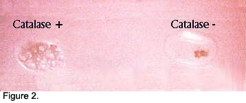
Special Features -
catalase is found in most aerobic and facultative anaerobic bacteria. The main
exception is Streptococci, which have catalase negative
test. Catalase is not found in anaerobes.
Precautions in interpretation:
It is important not to contaminate the bacterial colony under test with blood
agar. Red blood cells contain catalase and their presence will give a false positive
result. Old cultures may loose their catalase activity, possible resulting in a
false negative result.
|
Microorganism |
Catalase test |
|
Staphylococci |
Positive (exception: Staphylococcus aureus, subspecies anaerobius is catalase negative) |
|
Streptococci |
Negative |
|
Actinomyces israelii |
Negative |
|
Fusobacterium |
Negative |
|
Prevotella melaninogenica |
Negative |
|
Family of Enterobacteriaceae |
Positive |
|
- Escherichia colli |
Rare types from moderate to mostly non-reactive |
|
- Salmonella |
Moderate positive reaction |
|
- Shigella |
Mostly non-reactive |
|
Clostridium |
Negative |
As stated in Catalase Test as an Aid to the Identification of
Enterobacteriaceae research paper
published in 1972, family of enterobacteriaceae have differentiation of
catalase positive reaction, from vigorous ( Serratia, Proteus and Providencia),
moderate ( Salmonella, some rare types of Escherichia, Enterobacter and
Klebsiela) to non-reactive (most types of Escherichia and Shigella).
___________________________________________________________________________________________
Bibliography:
Levinston W., Jawetz E.; Microbiology and Immunology, 4-th edition 1996
University of Kentucky: http://www.mc.uky.edu/oaa/curriculum/iid98/manual/00lab2.htm
University of Texas, Houston Medical School: http://medic.med.uth.tmc.edu/path/tests.htm
TAYLOR W.I. and ACHANZAR D.; Catalase Test as an Aid to the
Identification of
Enterobacteriaceae- 1972 American Society for Microbiology
The University of Newcastle, Australia: http://whitewolf.newcastle.edu.au/techinfo/proc_bacto_biochem.html
Microbionet: http://www.microbionet.com.au
_________________________________________________________________________________
|
|
 |
|
 |